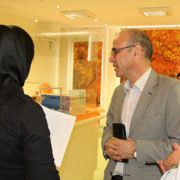
حضور مدیر عامل محترم شرکت در بخش اهدا مرکز جمع اوری پلاسمای فردیس
/۰ دیدگاه /در مقالات /توسط هادی حجت پناهعید سعید فطر بر شما مبارک
/۰ دیدگاه /در مقالات /توسط هادی حجت پناهIntravenous immunoglobulins
/۰ دیدگاه /در مقالات /توسط آقای زهفروشIntravenous immunoglobulins in liver transplant patients: Perspectives of clinical immune modulation
Arno Kornberg
Abstract
Shortage of appropriate donor grafts is the foremost current problem in organ transplantation. As a logical consequence, waiting times have extended and pretransplant mortality rates were significantly increasing. The implementation of a priority-based liver allocation system using the model of end-stage liver disease (MELD) score helped to reduce waiting list mortality in liver transplantation (LT). However, due to an escalating organ scarcity, pre-LT MELD scores have significantly increased and liver recipients became more complex in recent years. This has finally led to posttransplant decreasing survival rates, attributed mainly to elevated rates of infectious and immunologic complications. To meet this challenging development, an increasing number of extended criteria donor grafts are currently accepted, which may, however, aggravate the patients’ infectious and immunologic risk profiles. The administration of intravenous immunoglobulins (IVIg) is an established treatment in patients with immune deficiencies and other antibody-mediated diseases. In addition, IVIg was shown to be useful in treatment of several disorders caused by deterioration of the cellular immune system. It proved to be effective in preventing hyperacute rejection in highly sensitized kidney and heart transplants. In the liver transplant setting, the administration of specific Ig against hepatitis B virus is current standard in post-LT antiviral prophylaxis. The mechanisms of action of IVIg are complex and not fully understood. However, there is increasing experimental and clinical evidence that IVIg has an immuno-balancing impact by a combination of immuno-supporting and immuno-suppressive properties. It may be suggested that, especially in the context of a worsening organ shortage with all resulting clinical implications, liver transplant patients should benefit from immuno-regulatory capabilities of IVIg. In this review, perspectives of immune modulation by IVIg and impact on outcome in liver transplant patients are described.
Passively Administered
/۰ دیدگاه /در مقالات /توسط آقای زهفروشPassively Administered Pooled Human Immunoglobulins Exert IL-10 Dependent Anti-Inflammatory Effects that Protect against Fatal HSV Encephalitis
Chandran Ramakrishna,1 Alain N. S. Newo,1 Yueh-Wei Shen,1 and Edouard Cantin1,2,3,*
Mark L. Buller, Editor
Abstract
HSV-1 is the leading cause of sporadic encephalitis in humans. HSV infection of susceptible 129S6 mice results in fatal encephalitis (HSE) caused by massive inflammatory brainstem lesions comprising monocytes and neutrophils. During infection with pathogenic microorganisms or autoimmune disease, IgGs induce proinflammatory responses and recruit innate effector cells. In contrast, high dose intravenous immunoglobulins (IVIG) are an effective treatment for various autoimmune and inflammatory diseases because of potent anti-inflammatory effects stemming in part from sialylated IgGs (sIgG) present at 1–۳% in IVIG. We investigated the ability of IVIG to prevent fatal HSE when given 24 h post infection. We discovered a novel anti-inflammatory pathway mediated by low-dose IVIG that protected 129S6 mice from fatal HSE by modulating CNS inflammation independently of HSV specific antibodies or sIgG. IVIG suppressed CNS infiltration by pathogenic CD11b+ Ly6Chigh monocytes and inhibited their spontaneous degranulation in vitro. FcγRIIb expression was required for IVIG mediated suppression of CNS infiltration by CD45+ Ly6Clow monocytes but not for inhibiting development of Ly6Chigh monocytes. IVIG increased accumulation of T cells in the CNS, and the non-sIgG fraction induced a dramatic expansion of FoxP3+ CD4+ T regulatory cells (Tregs) and FoxP3− ICOS+ CD4+ T cells in peripheral lymphoid organs. Tregs purified from HSV infected IVIG treated, but not control, mice protected adoptively transferred mice from fatal HSE. IL-10, produced by the ICOS+ CD4+ T cells that accumulated in the CNS of IVIG treated, but not control mice, was essential for induction of protective anti-inflammatory responses. Our results significantly enhance understanding of IVIG’s anti-inflammatory and immunomodulatory capabilities by revealing a novel sIgG independent anti-inflammatory pathway responsible for induction of regulatory T cells that secrete the immunosuppressive cytokine IL-10 and further reveal the therapeutic potential of IVIG for treating viral induced inflammatory diseases.
IVIg Immune
/۰ دیدگاه /در مقالات /توسط آقای زهفروشIVIg Immune Reconstitution Treatment Alleviates the State of Persistent Immune Activation and Suppressed CD4 T Cell Counts in CVID
Dominic Paquin-Proulx,#1 Bianca A. N. Santos,#2 Karina I. Carvalho,2 Myrthes Toledo-Barros,2 Ana Karolina Barreto de Oliveira,2 Cristina M. Kokron,2 Jorge Kalil,2 Markus Moll,1 Esper G. Kallas,2, and Johan K. Sandberg1,*
Derya Unutmaz, Editor
Abstract
Common variable immunodeficiency (CVID) is characterized by defective B cell function, impaired antibody production, and increased susceptibility to bacterial infections. Here, we addressed the hypothesis that poor antibody-mediated immune control of infections may result in substantial perturbations in the T cell compartment. Newly diagnosed CVID patients were sampled before, and 6–۱۲ months after, initiation of intravenous immunoglobulin (IVIg) therapy. Treatment-naïve CVID patients displayed suppressed CD4 T cell counts and myeloid dendritic cell (mDC) levels, as well as high levels of immune activation in CD8 T cells, CD4 T cells, and invariant natural killer T (iNKT) cells. Expression of co-stimulatory receptors CD80 and CD83 was elevated in mDCs and correlated with T cell activation. Levels of both FoxP3+ T regulatory (Treg) cells and iNKT cells were low, whereas soluble CD14 (sCD14), indicative of monocyte activation, was elevated. Importantly, immune reconstitution treatment with IVIg partially restored the CD4 T cell and mDC compartments. Treatment furthermore reduced the levels of CD8 T cell activation and mDC activation, whereas levels of Treg cells and iNKT cells remained low. Thus, primary deficiency in humoral immunity with impaired control of microbial infections is associated with significant pathological changes in cell-mediated immunity. Furthermore, therapeutic enhancement of humoral immunity with IVIg infusions alleviates several of these defects, indicating a relationship between poor antibody-mediated immune control of infections and the occurrence of abnormalities in the T cell and mDC compartments. These findings help our understanding of the immunopathogenesis of primary immunodeficiency, as well as acquired immunodeficiency caused by HIV-1 infection.
Human Albumin
/۰ دیدگاه /در مقالات /توسط آقای زهفروشHuman Albumin Prevents 6-Hydroxydopamine-Induced Loss of Tyrosine Hydroxylase in In Vitro and In Vivo
Li-Juan Zhang,1,2 Yue-Qiang Xue,3 Chun Yang,1 Wei-Hua Yang,1 Long Chen,4 Qian-Jin Zhang,5 Ting-Yu Qu,6 Shile Huang,4 Li-Ru Zhao,7 Xiao-Min Wang,2,* and Wei-Ming Duan1,
Abstract
Human albumin has recently been demonstrated to protect brain neurons from injury in rat ischemic brain. However, there is no information available about whether human albumin can prevent loss of tyrosine hydroxylase (TH) expression of dopaminergic (DA) neurons induced by 6-hydroxydopamine (6-OHDA) toxicity that is most commonly used to create a rat model of Parkinson’s disease (PD). In the present study, two microliters of 1.25% human albumin were stereotaxically injected into the right striatum of rats one day before or 7 days after the 6-OHDA lesion in the same side. D-Amphetamine-induced rotational asymmetry was measured 7 days, 3 and 10 weeks after 6-OHDA lesion. We observed that intrastriatal administration of human albumin significantly reduced the degree of rotational asymmetry. The number of TH-immunoreactive neurons present in the substantia nigra was greater in 6-OHDA lesioned rats following human albumin-treatment than non-human albumin treatment. TH-immunoreactivity in the 6-OHDA-lesioned striatum was also significantly increased in the human albumin-treated rats. To examine the mechanisms underlying the effects of human albumin, we challenged PC12 cells with 6-OHDA as an in vitro model of PD. Incubation with human albumin prevented 6-OHDA-induced reduction of cell viability in PC12 cell cultures, as measured by MTT assay. Furthermore, human albumin reduced 6-OHDA-induced formation of reactive oxygen species (ROS) and apoptosis in cultured PC12 cells, as assessed by flow cytometry. Western blot analysis showed that human albumin inhibited 6-OHDA-induced activation of JNK, c-Jun, ERK, and p38 mitogen-activated protein kinases (MAPK) signaling in PC12 cultures challenged with 6-OHDA. Human albumin may protect against 6-OHDA toxicity by influencing MAPK pathway followed by anti-ROS formation and anti-apoptosis.
Contact Activation of Blood Plasma and Factor XII
/۰ دیدگاه /در مقالات /توسط آقای زهفروشContact Activation of Blood Plasma and Factor XII by Ion-exchange Resins
Chyi-Huey Josh Yeh, Ziad O. Dimachkie, Avantika Golas, Alice Cheng, Purnendu Parhi and Erwin A. Vogler*
A Contribution from the Hematology at Biomaterial Interfaces Research Group
Abstract
Sepharose ion-exchange particles bearing strong Lewis acid/base functional groups (sulfopropyl, carboxymethyl, quarternary ammonium, dimethyl aminoethyl, and iminodiacetic acid) exhibiting high plasma protein adsorbent capacities are shown to be more efficient activators of blood factor XII in neat-buffer solution than either hydrophilic clean-glass particles or hydrophobic octyl sepharose particles (a.k.a autoactivation, where FXII is the zymogen and FXIIa is a procoagulant protease). In sharp contrast to the clean-glass standard of comparison, ion-exchange activators are shown to be inefficient activators of blood plasma coagulation. These contrasting activation properties are proposed to be due to the moderating effect of plasma-protein adsorption on plasma coagulation. Efficient adsorption of blood plasma proteins unrelated to the coagulation cascade impedes FXII contacts with ion-exchange particles immersed in plasma, reducing autoactivation, and causing sluggish plasma coagulation. By contrast, plasma proteins do not adsorb to hydrophilic clean glass and efficient autoactivation leads directly to efficient activation of plasma coagulation. It is also shown that competitive-protein adsorption can displace FXIIa adsorbed to the surface of ion-exchange resins. As a consequence of highly-efficient autoactivation and FXIIa displacement by plasma proteins, ion-exchange particles are slightly more efficient activators of plasma coagulation than hydrophobic octyl sepharose particles that do not bear strong Lewis acid/base surface functionalities but to which plasma proteins adsorb efficiently. Plasma proteins thus play a dual role in moderating contact activation of the plasma coagulation cascade. The principal role is impeding FXII contact with activating surfaces but this same effect can displace FXIIa from an activating surface into solution where the protease can potentiate subsequent steps of the plasma coagulation cascade.
اطلاعات تماس
تهران، بزرگراه شیخ فضل الله نوری،تقاطع شهید همت،جنب برج میلاد
تلفنخانه: ۴۳۶۹۰۰۰۰-۰۲۱
دورنگار: ۸۸۶۲۹۱۲۰-۰۲۱


